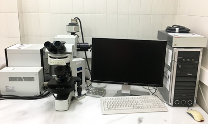
1) The Confocal Laser Scanning Microscope Leica TCS SP8 unit is equipped with one inverted microscope (model DMI6000 CS) with multiple DICM and fluorescent features-capabilities. Oil-water immersion and dry objective lenses are fully motorized and attached to the microscope unit. Attached to the main microscope, a high-end Scientific fluorescence CCD camera (model Leica DFC365FX) is available for executing fast, real-time recording. The system has an antivibration stage, an advanced and high-speed Laser Scanning System Unit, and it is also equipped with a set of high-quality lasers (UV 405nm Diode, DPSS 561nm, He-Ne 633nm, Argon 458-476-488-496-514nm).
The state-of-the-art computer system is capable—through the powerful software LAS AF 3 (Leica Application Suite Advanced Fluorescence)—of acquisition and analysis of the data. It offers full control over the microscope hardware and provides all necessary information. LAS AF 3 is fully synchronized with the programmable control panel, which allows interactive and fast setup.
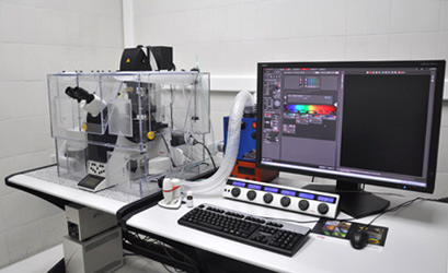
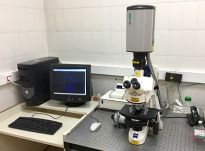
2) The Zeiss-Biorad unit is equipped with 1 upright microscope with DIC and fluorescent capabilities and oil-immersion lenses, 1 Vibration isolation table, and the Laser Scanning System Unit. The microscope unit is manufactured by Carl Zeiss company (model: Axioskop 2 Plus) and a Scanning Head is attached on it. The Laser Scanning System is manufactured by Biorad company (model: Radiance 2100 equipped with 3 lasers (Argon, He-Ne, Red Iod). The Facility is also equipped with a computer system (hardware plus software) for data acquisition and analysis.
The Zeiss-Biorad Laser scanning confocal microscope unit (below)
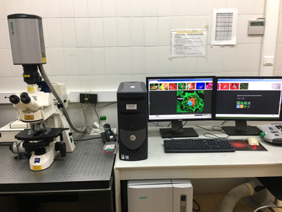
3) The BX61 microscope system incorporates a motorized Z focus that interfaces with a laser-based autofocus unit for active focus tracking, making inspections faster and more easily reproducible.
The Olympus unit BX61 is a fully motorized research microscope built on the platform of the traditional upright frame. In the system, a DSU (Disk Scanning Unit) spinning disk confocal is integrated that contains a pattern of slits that creates a virtual pinhole as the disk spins. All major microscope functions are completely automated, including focus, illumination, objective lens selection, and filter wheels. All motorized accessories are driven by a master external system controller, the BX-UCB. The unit is also equipped with a state-of-the-art computer system for data acquisition and analysis. A complete software (Cell-R) command set allows for full computer control.